Accidental surgical treatment of ankle injuries


- Have you just started exploring treatment options, or has your GP referred you? You're in the right place! We can help you get a complete overview of the surgical and non-surgical solutions for ankle injuries.
Injuries to the ankle
Ankle injuries are one of the most common traumatological conditions. If a bad step, jump or fall causes the foot to twist and the ankle joint to turn inwards or outwards, it can lead to ligament damage or even an ankle fracture. Professional, accident-surgical treatment is essential for a speedy and complete recovery, as without treatment, permanent damage to the joint, the surrounding ligamentous system and the joint surfaces can occur.
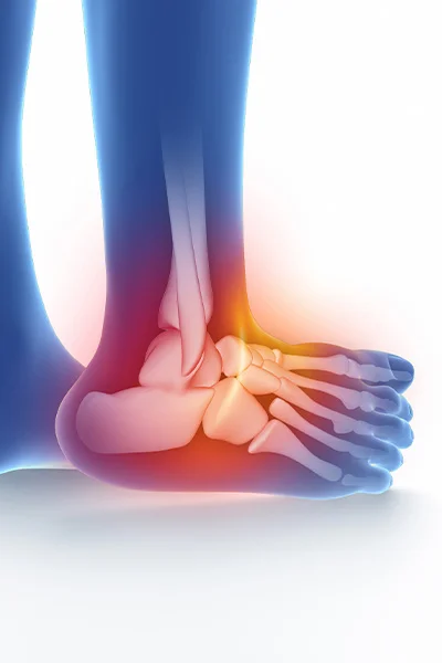
Anatomy of the ankle joint
The ankle joint is made up of 3 bones:
- The lateral malleolus: the distal end of the fibula
- The inner ankle: the distal end of the tibia
- The maxillary talus
The bones are tightly interconnected by ligaments that ensure the stability of the joint. In addition to the ligamentous system of the outer and inner ankle, the tibia and fibula are also connected by a tight ligamentous system (syndesmosis).
Healthy taste surfaces are covered with glass cartilage that slides easily over each other, lubricated by synovial fluid that also nourishes the articular cartilage.
If the ankle joint is unstable, it can speed up the process of joint wear.
The most common causes and types of ankle injuries
Causes of ankle injuries
- Injuries following a wrong step
- Injuries following stamping or slipping of the foot
- Injuries following a fall
- Twisting, entrapment injuries following a car accident
Types of ankle injuries
Tears
The ligament system around the ankle is torn and cannot perform its function.
Significant oedema, swelling and haematoma may develop around the patient's ankle, and the patient's gait becomes difficult and unsteady.
Sprains
The ligamentous system around the ankle is stretched and stretched. The ligament system does not tear completely, but it can swell painfully.
Breakages
A fracture with or without displacement (crack) or with displacement in one of the bones that make up the ankle joint. The affected joint becomes swollen and painful.
- The affected joint becomes swollen and painful.
- Moderate to significant deformity is seen around the ankle.
- Smaller or larger blisters often appear on the skin.
- The step becomes difficult or the patient becomes unable to walk.
Post-injury investigation
Following an ankle injury, the doctor will lie on the examination table with the knee in a flexed position and examine first the healthy, intact limb, then the injured limb, including the leg and foot as well as the painful parts.
Following the physical examination, the doctor will request an X-ray and/or ultrasound scan at his or her discretion. The X-ray examination is usually a 3-way ankle scan, which is supplemented by an X-ray of the leg and foot if necessary. Ultrasound is also used to examine the soft tissues and ligaments.
If there is a penetrating injury to the upper humerus, a CT scan is usually performed. If there is a possibility of damage to the articular cartilage, MR examination helps to make a more accurate diagnosis.
Care options after the investigation
After the imaging scan, the doctor will evaluate the results and explain to the patient what abnormalities or lesions he or she has found.
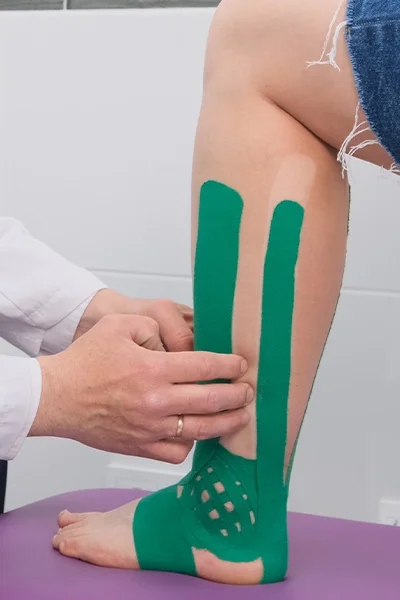
In the event of a sprain, if the ankle joint is stable and no major bleeding is detected, an elastic wrap is applied. In addition to resting the limb, the ankle injury heals quickly with the use of ice and topical creams to reduce the bleeding.
For ligament injuries and rupture of the joint capsule an orthosis, or anchorage, is applied. With an orthosis, up and down movement of the ankle is possible, but lateral twisting movements are prevented by the ankle brace. If worn for 5 weeks, the ankle injury will heal in the vast majority of cases. In the rare cases where the ankle remains unstable, it can be subsequently treated with ligament replacement.
In case of breakage surgery is usually the answer. The surgical treatment involves fitting the fractured ends together and fixing them with metal material (usually screws and plates). The aim of the surgery is to restore the anatomical structures and achieve a stable position in terms of movement, so in most cases no plaster fixation is needed.
For fractures without displacement, or if the patient has many other concomitant internal medical conditions and the fractured limbs are in an acceptable position in the cast applied after the fracture, conservative treatment, usually with a cast, may be attempted. This initially involves a recumbent cast and then, after a period of time, a walking cast.
BMM ankle doctors

Dr. György Kocsis PhD
Orthopaedic traumatologist, hand surgeon, assistant professor, wrist, elbow and shoulder surgeon, upper limb specialist

Dr. János Bartha
Orthopaedic-traumatologist, robotic surgeon
Plan your surgery: what to do before and after
You are about to undergo an acute operation, which is necessary to heal your fracture quickly and in the right position. The aim of the surgery is to restore anatomical structures and to achieve early joint motion. This will prevent the development of significant muscle atrophy or a severe restriction of joint movement.
We all want your ankle surgery to be as successful as possible. The following knowledge aims to improve the outcome of surgery, reduce the risk of complications and make the operation as safe and effective as possible.
Step 1
Be prepared, plan your surgery period!
Think about who will be your Helper during this period, who could be a close friend or family member. The Helper will be the person who:
- Accompanies you to pre-operative tests following an ankle injury
- On the day of surgery, you will be admitted to hospital
- Regularly visits
- Take home on the day of issue
- Helps you at home
- Replaces the medicines you need
- Support, encouragement and support during the weeks of recovery
The presence of a Facilitator will greatly assist your successful preparation before surgery and your rehabilitation after surgery. With your help, you can join the ranks of the recovered, pain-free.
Step 2
The last visit to the surgery
Once you and your doctor have decided on the surgery, you can start preparing for it.
- Obtaining imaging studies/x-rays: these are necessary for pre-operative planning. You will also have a recent X-ray taken at the hospital before the operation, which will be used by the surgeon to plan the operation.
- Laboratory tests, blood tests: these are done at the hospital where the operation is performed as part of the investigation.
- Specialist findings: during the pre-operative assessment, the anaesthetist will determine what previous or current specialist tests are required.
- If possible, the Facilitator should accompany you to your last office visit.
Step 3
Pre-operative hospital tests
Before the operation, laboratory tests will be carried out and, if the anaesthetist thinks it necessary, a chest X-ray will be taken.
If you are taking any anticoagulant medicines, be sure to tell your doctor because these medicines increase the risk of bleeding complications and should be discontinued or, if possible and necessary, suspended before surgery.
Step 4
The day of the operation
Depending on the nature of the fracture, the operating trauma surgeon performs a bone fusion operation using a plate, screw or staple and compression sling, or a combination of these.
During the surgery, he makes incisions above the outer ankle and/or the inner ankle, which are sutured shut at the end of the surgery.
At the end of the operation, the wound and the surgical site are infiltrated with painkillers and anti-bleeding injections, which significantly reduce the risk of bleeding complications and post-operative pain.
The doctor will sometimes leave a tube in the wound to drain the bleeding, which is removed the first day after surgery. After the operation, the wound is covered with a dressing and an elastic bandage is applied to the leg. If the surgeon deems it appropriate, a resting plaster splint is applied.
Step 5
Going inside, going home
After the operation, you can go home the same day or the first day after the operation, depending on the nature of the fracture and the procedure.
The next day after surgery, you will have a medical visit in the morning to have your wound checked. The wound will be covered with a waterproof dressing so you can shower without getting your dressing wet.
With the help of a physiotherapist, you can get up and walk with two elbow crutches to relieve the strain on the injured limb. You will be taught how to get around, get off and on the bed.
In the event of post-operative pain, nurses will give you a painkiller infusion or tablets.
On the day of return home, he or she is able to move, eat, drink and clean up independently. We will arrange your return home with you in advance.
In a normal passenger car, you can travel home sitting in the back seat with the injured limb in the back seat. When getting in and out of the car, your helper will assist you in sitting down and standing up.
Step 6
A few tips for home
- In his home, he shelves the injured limb
- Exercise your free joints
- Ice for 10-15 minutes every hour through a bandage
- You cannot put weight on the operated side, but you can walk with an aid
DO NOT FELEDJE! Regular practice of the physiotherapy exercises you have learned is one of the keys to a safe recovery. Exercise helps blood circulation, which is good for your hips and wound healing.
FREQUENTLY ASKED QUESTIONS
Suture removal is scheduled for 8-12 days after surgery. After the sutures have been removed, the wound can be flooded after 24 hours.
In the first few weeks, the surgeon may allow walking with partial weight-bearing after the external orthosis has been fitted, using 1 or 2 elbow crutches.
A follow-up X-ray of your injured ankle is taken in the 6th week after surgery. We can monitor the healing process with the help of the X-rays. If it is progressing well, the bracing treatment used can be abandoned.
At the same time, it is recommended to start intensive physiotherapy to regain the restricted joint movements and the lost muscles.
You should have a follow-up X-ray 3 months and 6 months after the operation.
Full bony healing takes six months.
Afterwards, if your surgeon deems it necessary, the previously inserted metal materials can be removed.
Yes, it is necessary. After lower limb surgery, an anticoagulant is injected under the skin until the weight-bearing operation to prevent clotting.
The aim of the surgery is to achieve a stable bone fusion. In some cases, the surgeon may wish to apply a plaster cast for a few days to protect the soft tissues. If a stable position is not achieved during the operation, additional post-operative plaster casts may be needed, the length and type of which will be decided by your doctor.
Not all ankle fractures require surgical treatment. Non-displaced fractures can be treated conservatively, with cast fixation or orthotic treatment.
In addition, if the patient has many internal medical conditions, the operation carries more risks than benefits, and an acceptable situation is found on follow-up X-ray after the reduction, fracture treatment with plaster fixation may be attempted.
The condition of the soft tissues was required because rest or surgery failed to achieve a stable position for movement, or movement may risk displacement of the fracture.
In young patients, it is advisable to remove the inserted metallic material at the earliest six months after surgery. In older patients with multiple underlying conditions, it may not be necessary to remove the inserted metal material.
Fractures that are displaced or prone to displacement require surgical treatment. If this is not done, there is a risk that the fracture will heal in the wrong position or that a false joint will develop, which can lead to painful ankle joints, difficulty walking or premature joint wear.
Fractures around the ankle result in interstitial swelling and haemorrhage. The increased tissue pressure can lead to small to large blisters on the skin. These make surgical treatment more difficult because they increase the risk of wound healing and infection.
In such cases, the surgeon will probably decide to postpone the operation and keep the limb in a cast until then.
The most common method of anaesthesia for the treatment of ankle fractures is spinal anaesthesia. An anaesthetic is administered at the level of the patient's lumbar spine, which numbs both lower limbs from the waist down, lasting 2-3 hours. During this time the patient feels no pain.
The time it takes to return to work depends to a large extent on the workplace and working conditions. A sedentary worker in an office can start working after the 3rd week, if access to work is arranged. If your work requires a long-term job or a lot of physical activity, it is not recommended until 2 months after surgery.
Yes, because the surgery reduces ankle movement on the injured side. Physical therapy is necessary to restore function and regenerate the muscles.
After the operation, it is advisable to keep the limb on ice for a week, and to relieve the weight. After this, if your doctor considers it appropriate, you can start with partial weight-bearing using a brace and then gradually reach full weight-bearing.
4 weeks after the operation, you can use your operated limb with a brace and full body weight.
It depends on the damaged side and your car. In the case of a left side fracture, if you have an automatic gearbox, you can drive for a week after the operation. In the case of a right side fracture or if you have a manual gearbox, you should only drive after 4 weeks after the operation.
Yes. Metal detectors usually alert you when a person with metal material (plate) passes through them. It is recommended that you tell security in advance, who will usually check with a metal detector. You do not need to bring a certificate with you, as it is not accepted by security services anywhere.
If you have any questions about ankle injuries, feel free to contact us!
CALL US NOW
Online booking